
Fig. 3.
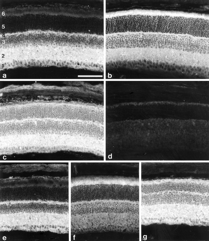
Immunohistological analysis of β2/β1+/+ and β2/β1ki/kimice. Immunohistological localization of β1 (a, c) and β2 (b) in sections of 17-d-old β2/β1+/+ (a, b) or β2/β1ki/ki (c) retinae using monoclonal antibodies BSP/3 (a, c) and 426 (b) recognizing β1 and β2 isoforms, respectively. Note the intense β1 immunoreactivity of inner segments of photoreceptor cells of β2/β1ki/ki mice. No β2 immunoreactivity is detectable on sections from β2/β1ki/ki mice incubated with monoclonal antibody 426 (d). The immunohistological localization of β1 (e, g) and β2 (f) in sections of 4-month-old β2/β1+/+ (e, f) or β2/β1ki/ki (g) retinae using monoclonal antibody BSP/3 (e, g) or 426 (f). Note the expression of β1 by photoreceptor cells of β2/β1ki/ki mice (g). 1, Ganglion cell layer and nerve fiber layer; 2, inner plexiform layer;3, inner nuclear layer; 4, outer plexiform layer; 5, outer nuclear layer;6, inner and outer segments of photoreceptor cells. Scale bar (shown in a for a–g): 100 μm.