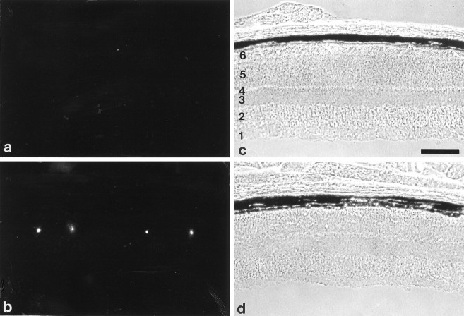
Fig. 7.
Apoptotic cell death of photoreceptor cells in 4-month-old β2/β1+/+ and β2/β1ki/ki mice. Visualization of apoptotic cell death in the retina of 4-month-old β2/β1+/+(a) and β2/β1ki/ki(b) mice using the TUNEL method. In the retina of 4-month-old β2/β1+/+ animals, apoptotic photoreceptor cells are virtually absent (a), whereas in retinae of 4-month-old β2/β1ki/kimice (b), apoptotic cell death of photoreceptor cells is frequently observed. c and drepresent the phase-contrast photomicrographs of a andb, respectively. 1, Ganglion cell layer and nerve fiber layer; 2, inner plexiform layer;3, inner nuclear layer; 4, outer plexiform layer; 5, outer nuclear layer;6, inner and outer segments of photoreceptor cells. Scale bar (shown in c for a–d): 100 μm.

