
Fig. 2.
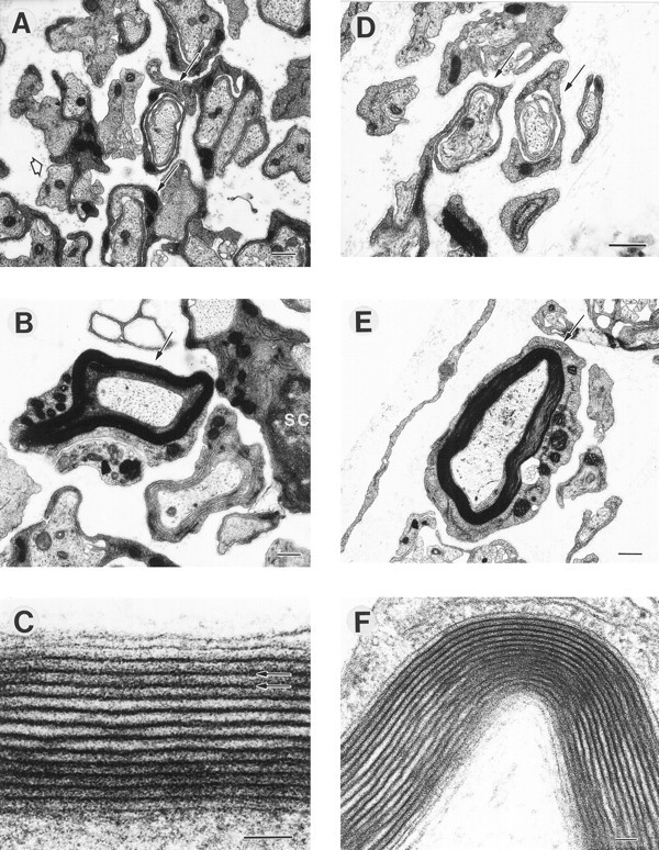
The ultrastructure of myelinated axons was normal in cultures stimulated at 0.1 Hz (A–C) and 1 Hz (D–F), and cultures under both stimulus conditions showed axons undergoing several stages of myelination. Note several loose wraps of Schwann cell cytoplasm ensheathing axons (A, D, arrows). Higher magnification of a myelinated axon is shown from cultures stimulated at 0.1 Hz (C, arrow) and 1 Hz (F, arrow). Multiple layers (up to 19) of compact myelin, with ∼10 nm between major dense lines (C, arrows), were evident in myelinated axons stimulated at 0.1 Hz (C) and 1 Hz (F). Scale bars: A, B, E, 500 nm; C, F, 50 nm.