
Fig. 6.
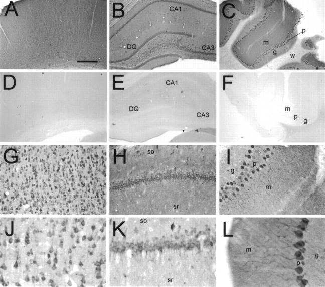
Immunohistochemical localization of yotiao in rat brain. Coronal sections were immunostained with yotiao antibodies B5843 (A, C–F,H, I, L) or B5844 (B, G, J,K) and visualized by DAB. These antibodies give similar staining patterns. A–C, Yotiao immunoreactivity in cerebral cortex (A), hippocampal formation (B), and cerebellum (C), with corresponding controls in D–F (antibody preincubated with immunogen). Note widespread somatodendritic labeling of neurons and diffuse staining of neuropil in neocortex, dentate gyrus, CA1 and CA3 of hippocampal formation, and cerebellum.G, J, Immunostaining of cell soma and apical dendrites of pyramidal neurons in the cerebral cortex. Staining of basal dendrites is also observed. H,K, Labeling of CA1 pyramidal cell layer in a somatodendritic manner. Note scattered interneuron immunoreactivity and neuropil staining in stratum oriens and stratum radiatum.I, L, Staining of Purkinje neurons extending from cell soma throughout major dendritic arborizations.DG, Dentate gyrus; m, molecular layer;g, granular layer; p, Purkinje cell layer; w, white matter; so, stratum oriens; sr, stratum radiatum. Scale bars:A–F, 0.5 mm; G–I, 125 μm;J–L, 62.5 μm.