
Fig. 7.
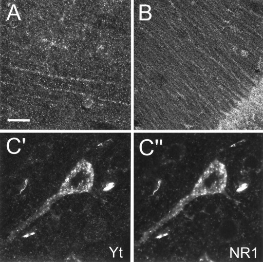
Localization of yotiao in rat cerebral cortex and hippocampus, and colocalization of yotiao and NR1 in pyramidal neurons, by confocal immunofluorescence microscopy.A, B, Specific labeling of distal dendrites of pyramidal cells and the surrounding neuropil in the rat cerebral cortex (A) and the CA1 region of the rat hippocampus (B) in a punctate pattern suggestive of synaptic localization. Brain sections were lightly digested with protease before immunohistochemical processing to enhance distal dendritic and neuropil staining. C′, C", Colocalization of yotiao (C′) and NR1 (C") in immunoreactive puncta in the cell soma and proximal apical dendrites of cortical pyramidal neurons. Brain sections were first stained for yotiao (B5843 visualized by a Cy3-conjugated secondary antibody) followed by immunolabeling for NR1 (54.1 visualized by tyramide amplification system, TSA-green). Scale bar:A, C′, C", 30 μm;B, 75 μm.