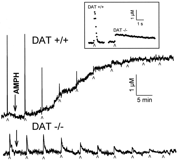
Fig. 1.
Effect of AMPH (10 μm) on DA efflux in striatal slices from wild-type (DAT +/+) and homozygote DAT knockout (DAT −/−) mice. Current was measured by cyclic voltammetry at microelectrodes implanted in the slices. Stimulated DA release was elicited by single electrical pulses applied to the slice at the times indicated by small arrowheads. On the time scale shown, electrically stimulated DA release events appear as sharp spikes. Baseline release was monitored as any increase in the current measured between stimulations that was identified as DA. In the absence of pharmacological or electrical intervention, baseline and electrically stimulated DA recordings were stable for >3 hr. Inset, Single-pulse stimulations in slices from a wild-type and homozygote DAT knockout mouse with the time scale expanded to show the time course of the DA release and clearance. Filled circles are individual measurements of the concentration of DA, collected every 100 msec.

