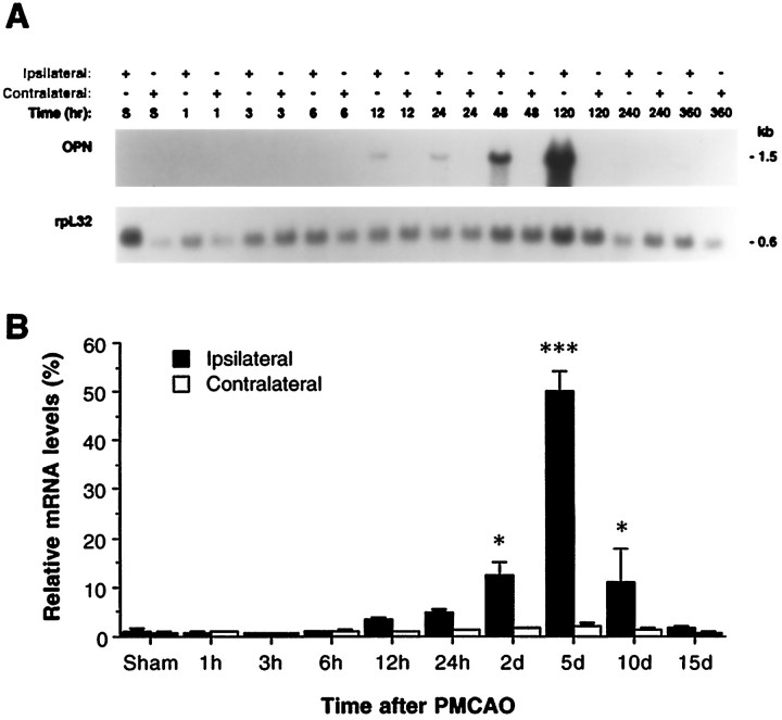
Fig. 1.
Northern blot analysis of OPN mRNA expression in rat ischemic cortex after permanent MCAO. A, Represent Northern blot for OPN and rpL32 cDNA probes to the samples from spontaneously hypertensive rats (SHR) after permanent MCAO. Total cellular RNA (40 μg/lane) was resolved by electrophoresis, transferred to a nylon membrane, and hybridized to the indicated cDNA probe. Ipsilateral and contralateral cortex samples (denoted by +) from individual rats of sham surgery (S; 5 d) or after 1, 3, 6, 12, and 24 hr and 2, 5, 10, and 15 d of permanent MCAO are depicted. B, Quantitative Northern blot data (n = 4) for OPN mRNA expression after focal stroke. The data were generated by PhosphorImager analysis and were displayed graphically after being normalized with rpL32 mRNA signals. *p < 0.05; ***p < 0.001, as compared with sham-operated animals.

