
Fig. 5.
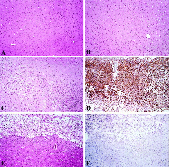
Histological and immunohistochemical staining of OPN expression in ischemic cortex. A, Hematoxylin and eosin-stained (H&E) section of normal cerebral cortex (sham-operated animals at day 5). B, H&E-stained focal ischemic zone at 24 hr after permanent MCAO. C, Focal ischemic zone at 5 d (H&E). D, Immunohistochemistry showing numerous OPN-positive macrophages in this 5 d lesion. E, Focal ischemic cortex at 15 d (H&E). F, Immunohistochemistry for OPN in focal ischemic lesion at 15 d.