
Fig. 3.
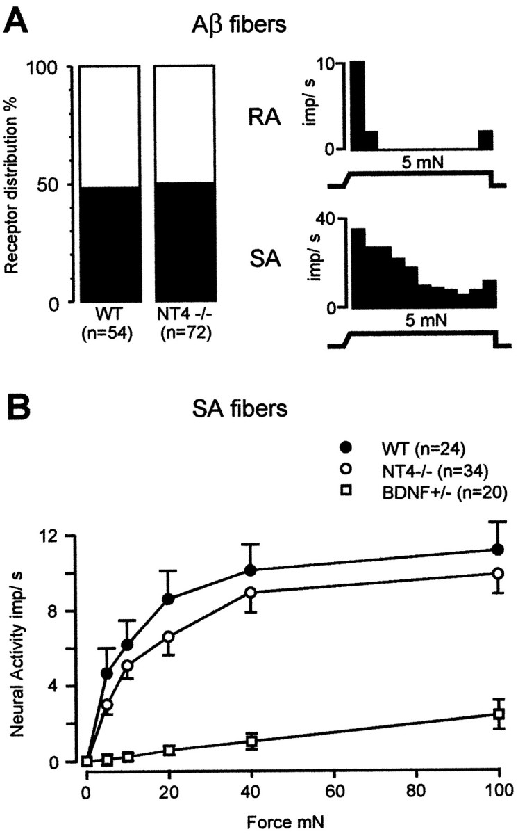
A, Left, Prevalence of subtypes of large myelinated (Aβ−) cutaneous fibers in WT and NT4−/− mice.Right, Representative examples of the mechanical response properties of an RA fiber (top) and an SA fiber (bottom). B, Stimulus–response functions of SA fibers to constant force stimuli. The stimulus–response functions of SA fibers in NT4−/− mice were not different from those of WT mice but differed strikingly from those found in BDNF-knock-out animals (BDNF-KO data from Carroll et al., 1998).