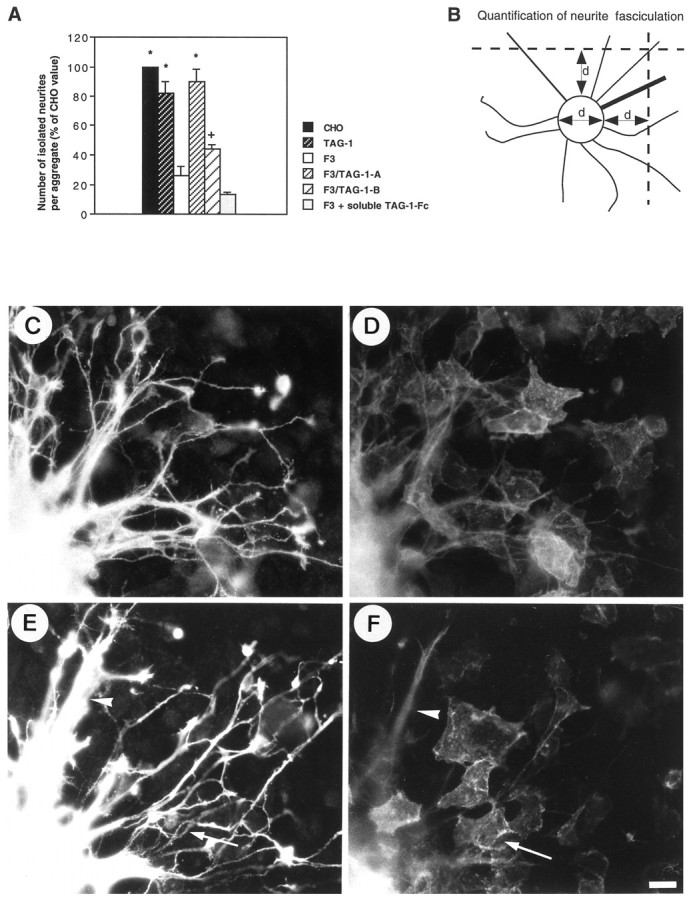
Fig. 6.

A, F3-mediated fasciculation of neurites from granule cell aggregates was completely prevented in the double-transfected F3-TAG-1-A CHO cells and only partly prevented in the F3-TAG-1-B clone. By contrast, soluble TAG-1-Fc added in the culture medium was unable to block neurite fasciculation induced by CHO-F3 cells. Measurements were taken on six granule cell aggregates under each experimental condition. Means are expressed as percentage of the mean control CHO value ± SEM. *Significant difference (p < 0.01); +significant difference (p < 0.05) with F3-CHO cells using ANOVA followed by Fischer’s PLSD test. B, Defasciculation of neurites was estimated as indicated in Material and Methods from the number of isolated neurites intersecting two transects drawn at one diameter (d) distance of the aggregate. The mean diameter of the (Figure legend continues) measured aggregates did not significantly vary under all the experimental conditions tested. C–F, Double-staining immunofluorescence of anti-GAP-43 rabbit antiserum (C, E) and anti-TAG-1 monoclonal antibody (D, F) in granule cell aggregates grown onto F3-TAG-1-A (C, D) and F3-TAG-1-B (E, F) monolayers. Note that neurites were defasciculated when grown on TAG-1-expressing cells (E, F, arrows), whereas bundles of neurites were observed on F3-only expressing cells (E, F, arrowheads). Scale bar, 10 μm.
