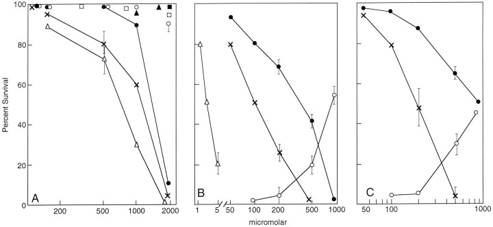
Fig. 1.
mGluR agonists and antagonists modify glutamate toxicity. Exponentially dividing cells were plated in 96-well plates as described in Materials and Methods, and 20 hr later the indicated reagents were added. Cell viability was determined by the MTT assay 24 hr later and verified by visual counting. All experiments were repeated at least three times with similar results. The error bars are the mean of triplicate assay points ± SEM. A, HT-22: ACPD, ×–×; glutamate, •–•; DHPG, ○–○; quisqualate, ▵–▵; U-73122, ▴–▴; AP-3, ■–■; and AIDA, ▪–▪.B, HT-22: AIDA, ×–×; AP-3, •–•; DHPG, ○–○; or U-73122 ▵–▵ were added to HT-22 cells 20 hr after plating at the indicated concentrations. Glutamate was added 30 min later at 1 mm to AIDA, U-73122, and AP-3 and at 1.5 mm to DHPG-containing cultures, and cell viability was determined by the MTT assay 24 hr later. Eighty-six percent of the cells survived in the presence of 1 mm glutamate alone. Because potentiation of toxicity was being assayed, the data were plotted with the 86% survival normalized to 100% survival for the sake of comparison with other data throughout the text. In the presence of the higher 1.5 mm glutamate concentration, only 51% of the cells survived. Because protection by DHPG was being assayed, 51% was normalized to zero (baseline) survival, and maximum protection (51–100%) was plotted as 100%. C, Cortical primary cultures: 2 d after plating, the culture medium was removed and replaced with fresh medium. To one set of cultures, AP-3 or AIDA was added at different concentrations followed by the addition of 2 mm glutamate. Viable cell number was determined 24 hr later by the MTT assay and verified by visual counting. In the absence of AIDA or AP-3, 81% of the cells survived. Cell death was potentiated by both AIDA (×–×) and AP-3 (•–•). Data are normalized to 81% survival, which is represented as 100% in the Figure. In another experiment, cells were exposed to increasing concentrations of DHPG (○–○) followed by 5 mm glutamate. In 5 mmglutamate alone, 47% of the cells survived. These data are normalized to 47% as 0% survival.

