
Fig. 2.
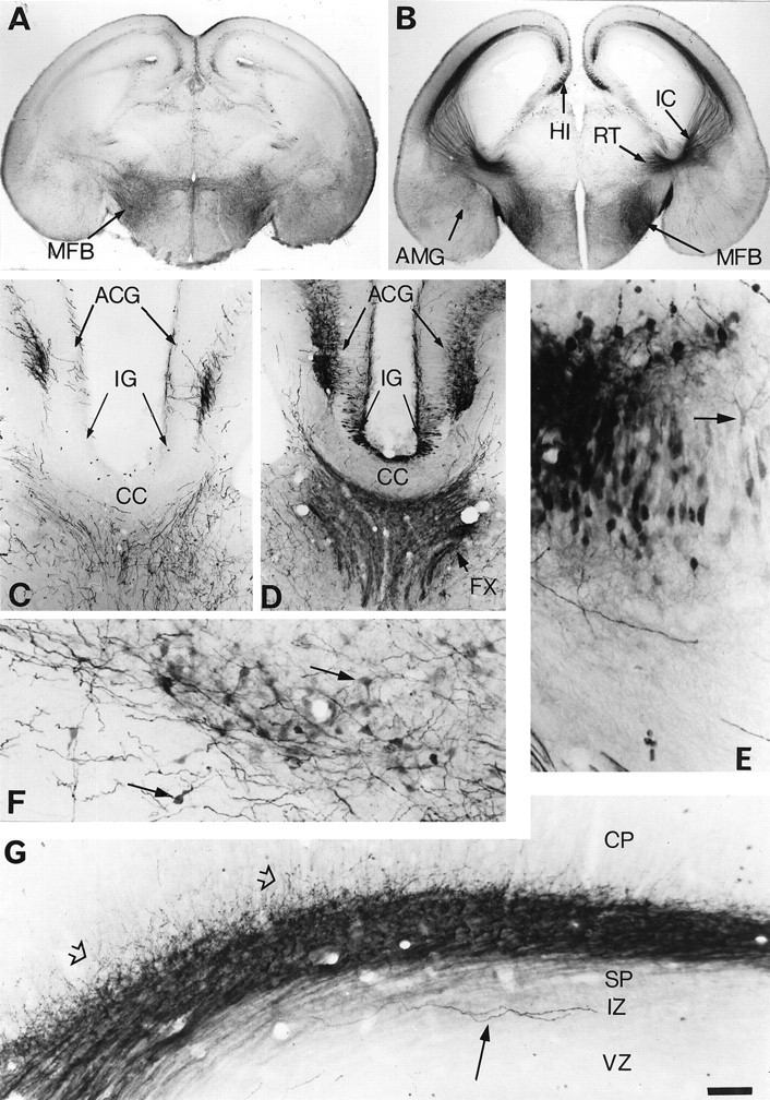
Atypical locations of 5-HT accumulation in the telencephalon of E18 MAOA knock-outs. Coronal brain sections are shown for controls (A, C) and MAOA knock-outs (B, D–G). A, In controls, 5-HT-immunostained fibers are primarily observed in the medial forebrain bundle (MFB). B, In MAOA knock-outs, 5-HT immunostaining of the MFB is increased, and a dense 5-HT immunolabeling is visible in the nucleus reticularis (RT), the thalamocortical fibers in the internal capsule (IC), the hippocampus (HI), and the amygdala (AMG). A higher magnification of the medial cortical area is shown inC and D at a more rostral level through the corpus callosum (CC), anterior cingulate cortex (ACG), and indusium griseum (IG).C, In controls, 5-HT immunoreactivity is only observed in terminal fibers or fiber tracts in the septum and ACG; the 5-HT-positive fibers in ACG form a bilaminar pattern in layer I and in the deep cortical layers. D, In MAOA knock-outs, 5-HT-positive fibers are more intensely stained, and additional labeling is visible in the fornix (FX) and in neuronal cell bodies in ACG and IG. E, A closer view of the 5-HT-immunolabeled cell bodies in the hippocampus reveals that these neurons have the morphological aspect of the principal pyramidal cells. Arrow indicates a neuron with a clear labeling of the dendritic tree. F, A closer view of the 5-HT immunolabeled neurons in the central nucleus of the amygdala.Arrows indicate neurons having a typical ovoid shape.G, Higher magnification of the 5-HT-positive thalamocortical fibers as they reach the cortical primordium. A dense network of fibers (fiber tracts and varicose fibers) is observed in the subplate (SP), with some fibers (open arrows) starting to penetrate in the cortical plate (CP). In contrast, a few long varicose fibers (arrow), probably representing afferents from the raphe, run in the intermediate zone (IZ). Only varicose fibers in SP and IZ were 5-HT immunoreactive in control mice, and this staining was much less intense than in MAOA knock-outs.VZ, Ventricular zone. Scale bar (in G):A, B, 625 μm; C,D, 150 μm; E, 27 μm;F, 40 μm; G, 33 μm.