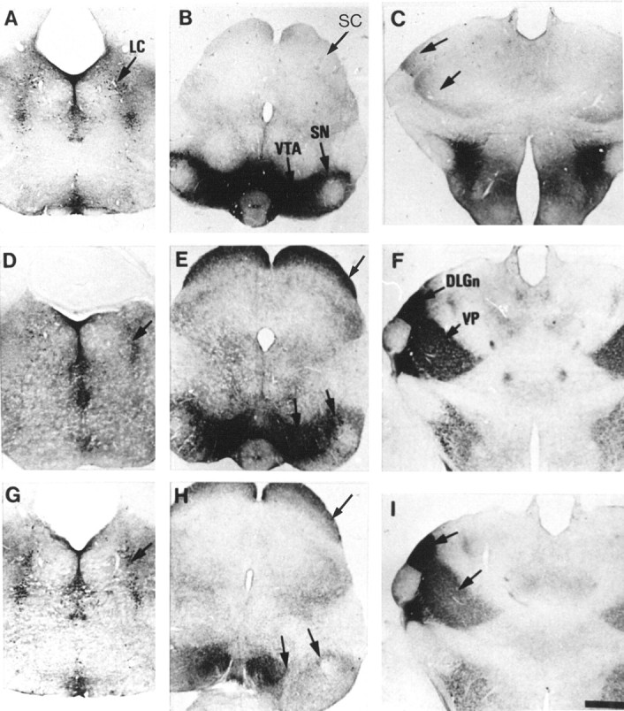
Fig. 6.

Changes of 5-HT immunoreactivity in P7 MAOA knock-outs after administration of selective inhibitors of monoaminergic transporters. Comparable coronal brain sections are shown in the metencephalon (A, D,G), mesencephalon (B, E,H), and diencephalon (C,F, I), after repeated administration of fluoxetine (A–C), nisoxetine (D–F), or GBR12783 (G–I) at P6 and P7. Control brain sections obtained from untreated MAOA knock-outs are not shown. 5-HT immunolabeling of the raphe nuclei is not visibly affected by any pharmacological treatment, although the staining of the fine varicose afferents from the raphe is reduced by the fluoxetine treatment. A–C, Fluoxetine, a selective inhibitor of SERT, causes the disappearance of 5-HT immunolabeling in the SC (B) and thalamus at the level of DLGn and VP (C) but increases staining of dopaminergic neurons in the SN and VTA (B), with no visible change in the LC (A). D–F, Nisoxetine, a selective inhibitor of NET, greatly reduces 5-HT immunolabeling in the LC (D) but does not cause changes of staining in the SN, VTA, SC (E), or thalamus (F). G–I, GBR12783, a selective inhibitor of DAT, abolishes 5-HT immunolabeling in the SN and VTA (H) but not in the LC (G), SC (H), or thalamus (I). Scale bar (inI): A–I, 625 μm.
