
Fig. 1.
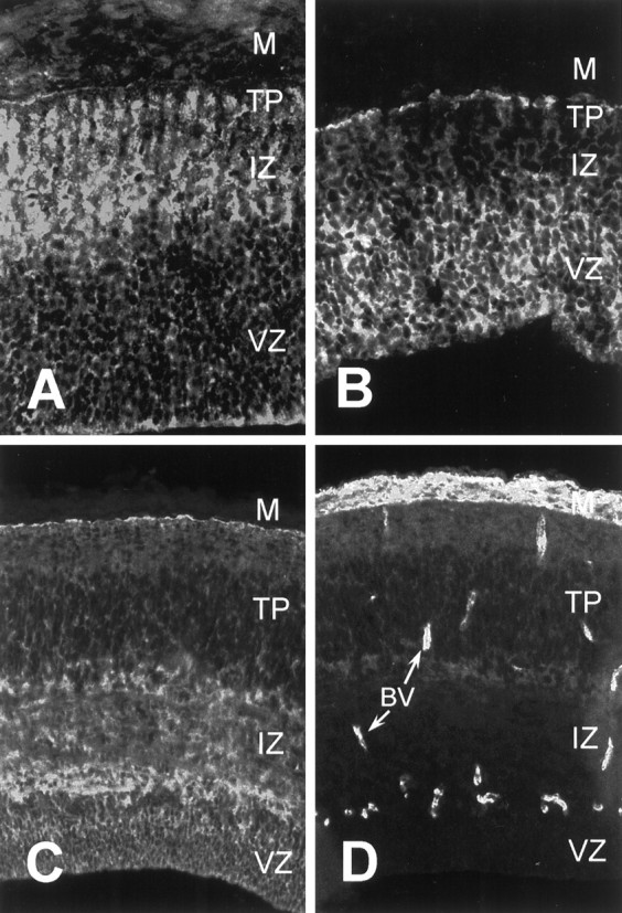
Expression of integrin subunits and substrates in the developing optic tectum. Cryosections of developing optic tectum were immunostained with antibodies against integrin α8 (A), integrin α6 (B), tenascin-C (C), and fibronectin (D). A, B, E7 tectum; C, D, E9 tectum. For all parts, the ventricular surface is down. Arrows inD denote blood vessels (BV).M, Meninges; VZ, ventricular zone;IZ, intermediate zone; TP, tectal plate. See Results for details.