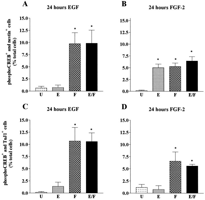
Fig. 8.
Characterization of growth factor-responsive cells in cultures of striatal precursor cells primed for 24 hr with EGF (A, C) or FGF-2 (B, D). Graphical representation of the percentage of double-immunopositive phospho-CREB and nestin (A, B) and phospho-CREB and TuJ1, (C, D) cells in unstimulated cultures (U) and in cultures stimulated with EGF (E), FGF-2 (F), or a combination of EGF and FGF-2 (E/F). Data represent the means of three independent experiments. For each condition a total of 600 cells were counted. A, B, *Significantly different from U(p < 0.01). C, D, *Significantly different from U(p < 0.001).

