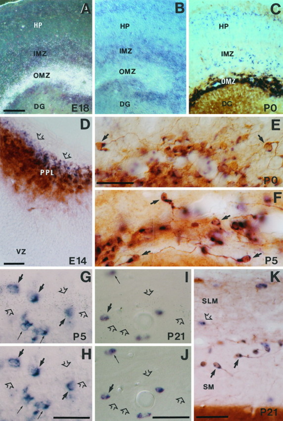
Fig. 5.

Characterization ofreelin-expressing cells in the hippocampal marginal zone–stratum lacunosum-moleculare at several developmental stages.A, B, Dark-field and bright-field photomicrographs of a double-labeled section (radioactive and nonradioactive ISH) showingreelin (silver grains inA) and GAD67 (blue in B) expression at E18; note the lack of colocalization of mRNAs in the outer marginal zone (OMZ). Weak reelinexpression is observed in the inner marginal zone (IMZ).C, Distribution of reelin mRNA (blue) and calretinin immunoreactivity (brown) in the OMZ and hippocampus at P0.D, Colocalization of reelin mRNA (blue) and calretinin immunoreactivity (brown) in the hippocampal preplate (PPL) at E14. Note the presence of cells expressing onlyreelin (open arrows) in the outer aspect of the preplate. E, F, Colocalization ofreelin mRNA (blue) and calretinin immunoreactivity (brown) in Cajal-Retzius cells (some are marked by bold arrows) near the hippocampal fissure at P0 (E) and P5 (F). Note the virtual complete colocalization of both labelings. G, H, Pair of photomicrographs taken at different planes of focus illustrating colocalization of reelin mRNA (silver grains, open arrows) and GAD67 expression (blue cells) in the stratum lacunosum-moleculare at P5. Although most reelin transcripts (open arrows) are outside GAD67-positive cells, some neurons colocalize both transcripts (bold arrows); thin arrows point to neurons expressing only GAD67 mRNA. I, J, Pair of photomicrographs at different planes of focus, showing colocalization of reelin mRNA (silver grains, open arrows) and GAD67 mRNA (blue cells) at P21 in the stratum lacunosum-moleculare near the hippocampal fissure; conventions as in G, H; note the presence ofreelin-positive/GAD67-negative neurons (open arrows). K, Distribution ofreelin mRNA (blue, open arrow) and calretinin immunoreactivity (brown) around the hippocampal fissure at P21; somereelin-expressing/calretinin-positive Cajal-Retzius cells are labeled by bold arrows; open arrow points to areelin-positive/calretinin-negative neuron.DG, Dentate gyrus; HP, hippocampal plate;IMZ, inner marginal zone; SLM, stratum lacunosum moleculare; SM, stratum moleculare;VZ, ventricular zone. Scale bars: A–C, 100 μm; D–K, 50 μm.
