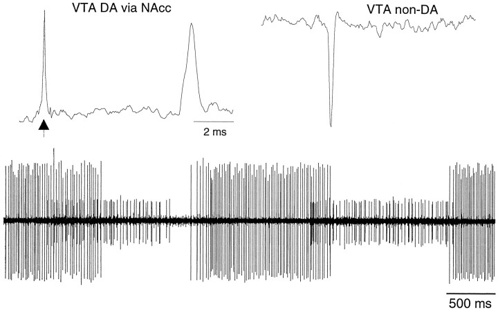
Fig. 1.
Extracellular electrophysiological characterization of dopamine and nondopamine neurons in the ventral tegmental area. Top, Unfiltered recordings of a VTA DA neuron evoked by stimulation of the NAcc (left) and of a spontaneous non-DA neuron (right) are shown. Calibration bar applies to both. VTA DA neurons were slow-firing (<1 Hz), bursting neurons that were driven by NAcc stimulation with spike durations of >500 μsec (arrow indicates NAcc stimulus artifact). VTA non-DA neurons were relatively fast-firing, nonbursting cells that evinced negative-going spikes and were characterized by spike durations of <500 μsec. VTA non-DA neurons were not driven by NAcc stimulation. Bottom, Under halothane anesthesia, VTA non-DA neurons evinced pronounced and persistent phasic activity as demonstrated by the two simultaneously recorded VTA non-DA neurons in the filtered trace.

