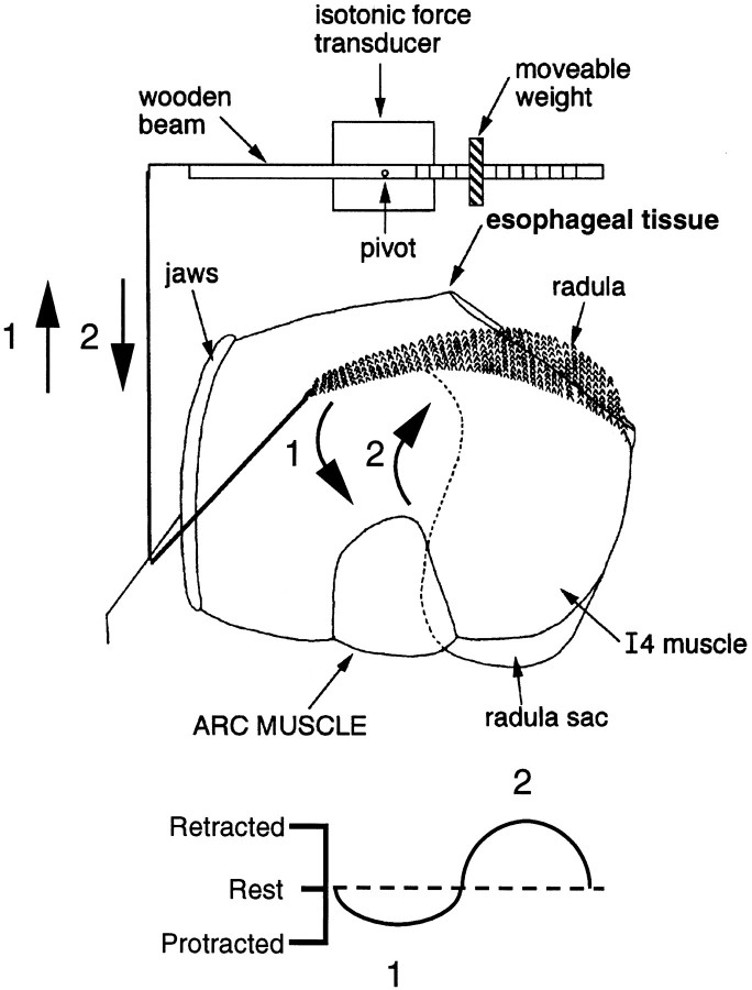
Fig. 1.
Schematic illustration of the preparation used to transduce movements of the buccal mass. (Ganglia are not shown.) A string was tied to the anterior tip of the radula and attached to a movement transducer. This transducer detects movement of the radula toward the jaws, which is referred to as protraction (arrow 1), and movement of the radula toward esophageal tissue, which is referred to as retraction (arrow 2). Protraction produces a downward deflection in transducer records, and retraction produces an upward movement. The rest position of the radula (the position before movement is elicited) is indicated by thedotted line in the bottom diagram.

