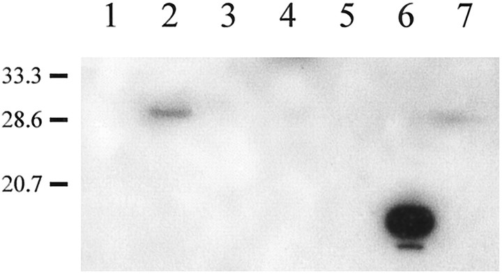
Fig. 5.
Western blot analysis of hybrid bass tissue homogenates probed with affinity-purified anti-Cx35 antisera.Lane 6 contains ∼1 μg of a crude lysate of the bacterial strain expressing the 6× His-tagged Cx35 intracellular loop peptide. Lanes 1 and 2 contain 10 μg of supernatant fractions of brain and retina, respectively. Ten micrograms of membrane pellets of the remaining tissues were loaded as follows: 3, heart; 4, spleen; 5, liver; 7, retina. Molecular weight markers are shown on the left. A 30 kDa band was strongly labeled in the retina supernatant fraction and was barely detectable in the 10,000 × g pellet.

