Fig. 6.

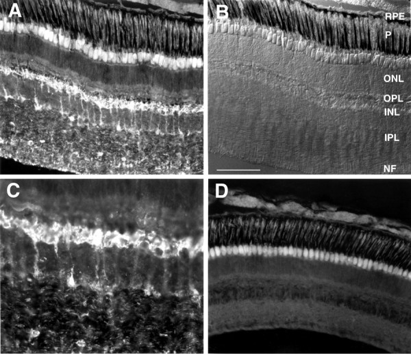
Immunofluorescent labeling of hybrid bass retina with affinity-purified anti-Cx35 antisera. A, Low-power micrograph of a transverse retinal section shows labeling of neurons in the inner nuclear, inner plexiform, and nerve fiber layers. No labeling was observed distal to the outer plexiform layer, nor were signals detected in the optic nerve (data not shown). B, Nomarski image of the field shown in A reveals the structure of the hybrid bass retina. RPE, Retinal pigment epithelium; P, photoreceptor layer;ONL, outer nuclear layer; OPL, outer plexiform (synaptic) layer; INL, inner nuclear layer;IPL, inner plexiform layer; NF, ganglion cell and nerve fiber layer. C, Higher-power view of the inner nuclear and inner plexiform layers from the section shown inA. Strongly stained cells in the inner nuclear layer appear to be bipolar cells. In addition, punctate labeling is present throughout much of the inner plexiform and ganglion cell layers; the cellular origin of this labeling is unclear. D, Control section lacking primary antibody shows the autofluorescence of the photoreceptor inner segments that was seen in A. Scale bar (in B): A, B,D, 100 μm; C, 50 μm.
