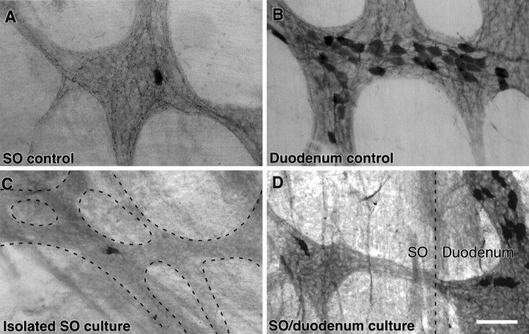
Fig. 5.
Calbindin-positive fibers in the SO degenerated when the SO was maintained in organ culture. A,B, Control preparations of the SO (A) and duodenal (B) ganglia that were fixed immediately and immunostained for calbindin and visualized with a DAB reaction product. Note the large number of calbindin-positive nerve fibers in the SO ganglia, relative to the single calbindin-positive neuron and the large number of calbindin-positive neurons in the duodenal ganglion. C, Photomicrograph of a region of the ganglionated plexus in the SO (dashed lines) that was immunostained for calbindin after the SO had been cultured in isolation for 72 hr. Note the absence of calbindin-positive nerve fibers. D, Photomicrograph of the interface between the SO and the duodenum (dashed line) in a preparation that was immunostained for calbindin after the SO had been cultured for 72 hr with the duodenum intact. Calbindin-immunoreactive nerve fibers can be seen in the interganglionic nerve bundle that passes between the duodenal and SO ganglia. Scale bar, 50 μm.

