
Fig. 2.
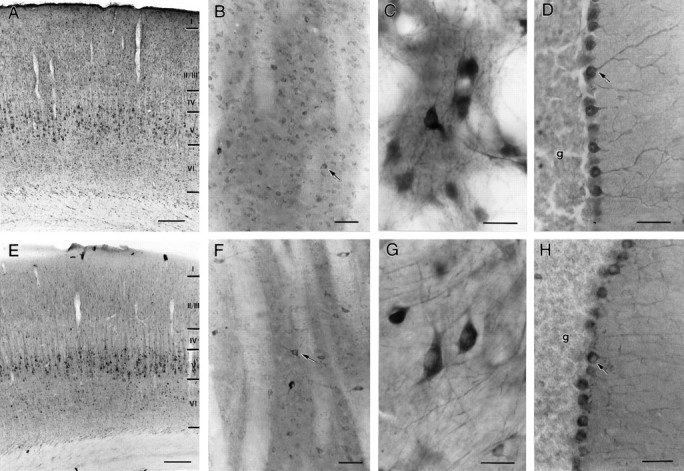
Similarities in HAP1 and huntingtin distribution in the rat brain. Light micrographs showing HAP1 (A–D) and huntingtin Hp549 (E–H) immunoreactivity are shown. Both HAP1 and huntingtin immunolabeling filled neuronal perikarya and dendrites. Throughout the brain, HAP1 cellular distribution was very similar to that of huntingtin; however HAP1 labeled more neurons than did huntingtin. A, E, In somatosensory cortex, HAP1 (A) and huntingtin (E) are found in pyramidal neurons.B, F, In striatum, both medium-sized neurons and larger neurons (arrows) are labeled with HAP1 (B) and huntingtin (F). C, G, In globus pallidus, neurons and dendrites are well stained by both HAP1 (C) and huntingtin (G).D, H, In the cerebellum, Purkinje cells (arrows) are more intensely stained by both HAP1 (D) and huntingtin (H) than are neurons in the granule cell layer (g). Scale bars: A,E, 200 μm; B, D,F, H, 50 μm; C,G, 25 μm.