
Fig. 5.
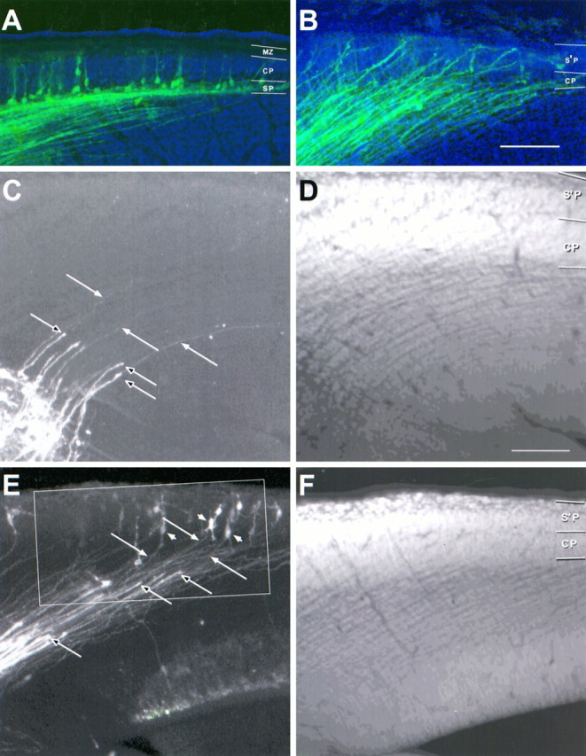
Here (and in Figs. 6, 7) the relationship among thalamocortical afferents, early corticofugal axons, and their cell bodies at E14.5 is seen in extended focus images of the cerebral wall of the left hemisphere made from thin optical sections collected by confocal microscopy (lateral is up; dorsal isright). A small crystal of DiI in the internal capsule has labeled a restricted group of both classes of axon; they appeargreen. The sections are counterstained with acridine orange; cell nuclei appear blue. The blue background is a bright-field image showing the boundaries of the cerebral wall.A, B, Low-power images of examples of wild-type (A) and reeler (B) mice in which thalamic fibers have already reached the cortex. The mass of axons, forming a parallel array in the intermediate zone, is a mixture of thalamocortical axons (many of which can be traced in high power to growth cones at their tips) and early corticofugal fibers (many of which can be followed back to the labeled cell bodies of origin. In the wild type (A), the labeled cells lie almost exclusively in the subplate (SP), with a very small number (some even with pyramidal morphology) in the cortical plate (CP) and, in other sections, in the marginal zone (MZ). In the reeler (B) no back-labeled somata are seen below the cortical plate (CP), through which both classes of axon run obliquely, up to the superplate (S’P) above, where labeled cell bodies are seen, specially at higher power (Fig. 7B). C, D and E, F, Examples from a reeler specimen from another E14.5 litter. Sections were cut at 45° to the coronal plane, approximately parallel to the labeled fibers over much of their course. The images on the left (C, E) show stained axons, whereas those on the right (D, F) show the acridine orange counterstaining, which reveals the characteristic loose cortical plate (CP) below the superplate (S’P).C, D, Close to the crystal placement in the internal capsule, a broad array of thick thalamic axons with growth cones at their tips (unfilled arrows) has advanced only a short distance into the intermediate zone. They are superimposed on an array of much finer axons, only just visible at this magnification (some indicated with filled arrows), extending back up toward the cortex. Many of these could be traced, in adjacent sections, to back-labeled cell bodies in the superplate. E, F, Further rostral, where development is more advanced, the thick thalamic axons have advanced further. The growth cones of some of them (unfilled arrows) are seen entering the cortical plate, clearly superimposed on a carpet of finer corticofugal axons (filled arrows), which can be followed back to labeled bipolar and multiform cell bodies in the superplate (examples indicated withfilled arrowheads). The region within the outline is shown at higher power in Figure 6. Confocal reconstructions: A, B, 32 sections × 2.0 μm intervals; D, 32 × 5.8 μm; E, 30 × 2.0 μm; D, 32 × 3.3 μm. Scale bars, 100 μm for all panels.