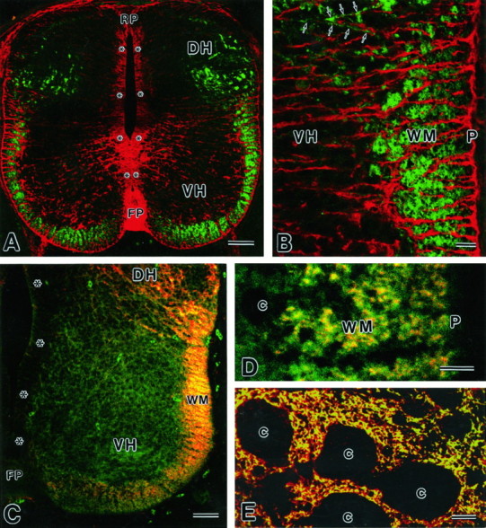
Fig. 4.

Images by confocal laser scanning microscopy.A, B, Double immunofluorescence for GLT-1 (green, rabbit antibody) and GLAST (red, guinea pig) in a paraffin cross-section at E13. GLT-1 immunoreactivity is seen in the marginal zone/white matter (WM) of the ventral cord and in the dorsal horn (DH), showing no overlaps with GLAST. A few transverse fibers (arrows) with GLT-1 immunoreactivity enter the ventral horn (VH). C,D, Double immunofluorescence for GLT-1 (red, guinea pig) and NSE (green, rabbit) in a cryostat cross-section at E13. Note their colocalization in the marginal zone/white matter (WM), but not in the ventral horn (VH). E, Double immunofluorescence for GLT-1 (green, rabbit) and GLAST (red, guinea pig) in a cross paraffin section at P14. c, Cell bodies; FP, floor plate; P, pia matter; RP, roof plate.Asterisks indicate the ventricular zone. Scale bars:A, C, 50 μm; B,D, E, 10 μm.
