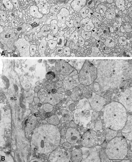
Fig. 5.

Electron micrographs of the spinal marginal zone at E13. A, Ultrastructure. Enlarged axonal portions are marked by asterisks.Arrowheads indicate growth cone parcels, which have been originally described for structures contained in growing pyramidal tract axons of neonatal rats (Gorgels, 1991a). B, Immunoelectron micrograph showing GLT-1. GLT-1 is detected in a small part of the axolemma apposing adjacent axons and sometimes to radial glial fibers (arrowheads). EF, End foot;RG, radial glial fiber. Scale bars, 1 μm.
