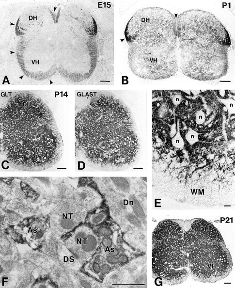
Fig. 6.

Spinal cords from E15 to P21. Cross paraffin sections were immunostained for GLT-1 (rabbit antibody) at E15 (A), P1 (B), P14 (C), and P21 (G), and for GLAST at P14 (rabbit antibody) (D).Arrowheads indicate GLT-1 immunoreactivity in the marginal zone/white matter. E, High-power view of the ventral cord in C. F, Immunoelectron micrograph at P14 showing the localization of GLT-1 in astrocytic processes (As) surrounding immunonegative nerve terminals (NT), dendritic spines (DS), and dendrites (Dn).DH, Dorsal horn; n, neuronal cell bodies;VH, ventral horn; WM, white matter. Scale bars: A–D, G, 100 μm;E, 10 μm; F, 1 μm.
