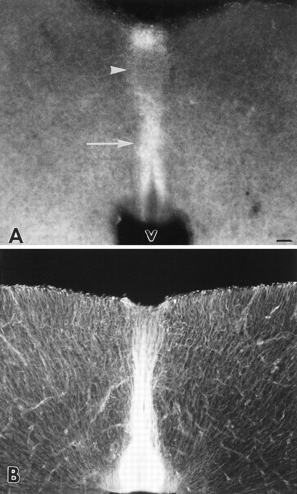
Fig. 1.

Immunostaining for CS and vimentin in coronal sections, 30 μm thick, through the tectum of the developing Syrian hamster at age P0. A, Section stained with monoclonal antibody CS-56, against chondroitin sulfate. Staining is concentrated in the midline region (arrow), in a band that extends between the ventricle and the pial surfaces. Note the lighter staining in the dorsal midline (arrowhead) where intertectal axons cross to the opposite side. V, Ventricle.B, Section stained with a monoclonal antibody against vimentin. Dense immunoreactivity is visible in midline cells, with more staining near the ventricle where the cell bodies of the midline glia lie. Scale bar, 50 μm.
