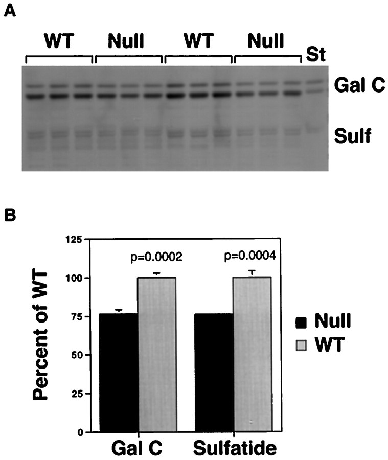
Fig. 4.
Myelin lipids in WT andIgf1−/− brains extracted from equal amounts of total protein demonstrated by thin layer chromatography. Orcinol-stained bands are identified as galactocerebroside (Gal C) or sulfatide (Sulf) by comparison of the comigrating standards (St) in A. The bands were quantified by the NIH Image analysis program, and the results are expressed as mean ± SEM of percentage of wild type (B).

