
Fig. 6.
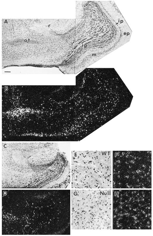
Oligodendrocyte concentration in olfactory bulb (A–D) and cerebellar medulla (E–H). PLP mRNA-positive cells are taken to be oligodendrocytes. In this figure, each bright-field micrograph is paired with a matching dark-field illumination showing the PLP mRNA-positive cells. The wild-type (WT) olfactory bulb (A, B) is much larger than that of the Igf1−/−(Null) (C, D). The number of mitral neurons and PLP mRNA-positive cells are both reduced in the Igf1−/−. ep, External plexiform layer; ig, internal granular layer;ip, internal plexiform layer; m, mitral cell layer; ot, olfactory tract. PLP mRNA-positive cells are shown at higher magnification on the cerebellar medulla of WT (E, F) andIgf1−/− (G,H) brain sections. Scale bar:A–D, 200 μm; E–H, 50 μm.