
Fig. 2.
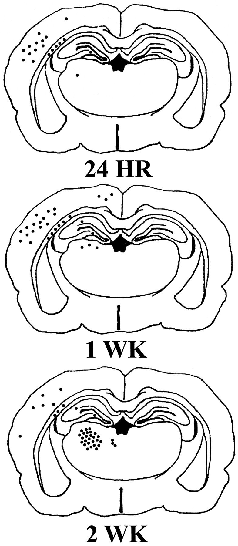
Schematic of TUNEL-stained sections of the rat brain 24 hr, 1 week, and 2 weeks after injury illustrating the regional distribution of apoptotic cells. Initially, apoptotic cells were observed primarily in the injured cortex at 24 hr after injury and over time were detected in internal structures, such as the thalamus and hippocampus. Each filled circle (•) represents five TUNEL-positive cells exhibiting apoptotic morphology.