
Fig. 4.
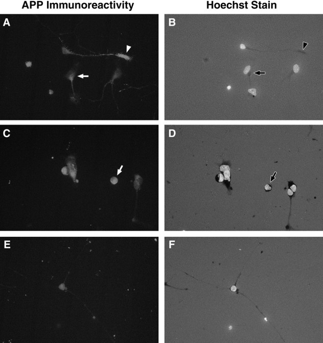
Immunocytochemistry shows increased expression of APP in motoneurons undergoing cell death in vitro. Motoneurons deprived of MEx (A, C) generally showed stronger APP immunoreactivity compared with those cultured with MEx (E). Aggregations of APP immunoreactivity were often observed in motoneurons in culture for 24 hr without MEx (A) in the cell body (arrow) or in the distal tip of the neurite (arrowhead). By 36 hr without MEx, many apoptotic motoneurons were observed in culture (arrow inD), and these cells were most often intensely APP-immunoreactive (arrow in C).A, C, E, APP immunoreactivity. B, D, E, The same field but with the UV filter to visualize the nuclei of the cells that were stained with the DNA-binding bis-benzimidazole dye Hoechst 33342. A, B, Motoneurons in culture for 24 hr without MEx. C, D, Motoneurons in culture for 36 hr without MEx. E,F, Motoneurons in culture for 36 hr with MEx. All fields are shown at the same magnification (40× objective). Representative photomicrographs are shown. Three independent cultures were observed, with two coverslips/condition/time point/culture.