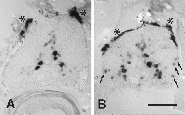
Fig. 12.
A, B, L1.2 mRNA was upregulated in glial cells in the spinal white matter caudal to the lesion site. All images are cross sections; dorsal is up. Asterisksindicate melanocytes covering the dorsal aspect of the spinal cord.A, Unlesioned control; B, 14 d post-lesion. Labeling of L1.2 mRNA was increased in small cells in the white matter (B, arrows) and in cells in the gray matter in the spinal cord caudal to a distal lesion (B) as compared with unlesioned controls (A). Scale bar (shown in B): 100 μm.

