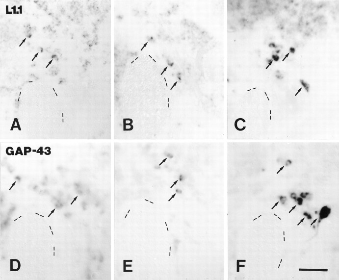
Fig. 8.
A–F, Expression of L1.1 and GAP-43 mRNAs in the nucleus of the lateral lemniscus was not significantly upregulated after distal but was upregulated after proximal lesion.In situ labeling of L1.1 (A–C) and GAP-43 (D–F) mRNAs, in unlesioned control fish (A, D), after distal (B, E) and proximal lesion (C, F) is shown. All images are cross sections; dorsal is up, lateral is left. Dashed lines outline the border of the lateral longitudinal fascicle as an anatomical landmark. Arrows point out individual cells in the nucleus of the lateral lemniscus. A–C, Labeling of L1.1 mRNA after distal lesion (B) was comparable to that in unlesioned controls (A), but it was strongly increased after proximal lesion (C). D–F, Intensity of labeling for GAP-43 mRNA was not increased after distal (E) but was increased after proximal lesion (F) as compared with unlesioned controls (D). Scale bar (shown in F forA–F): 50 μm.

