
Fig. 1.
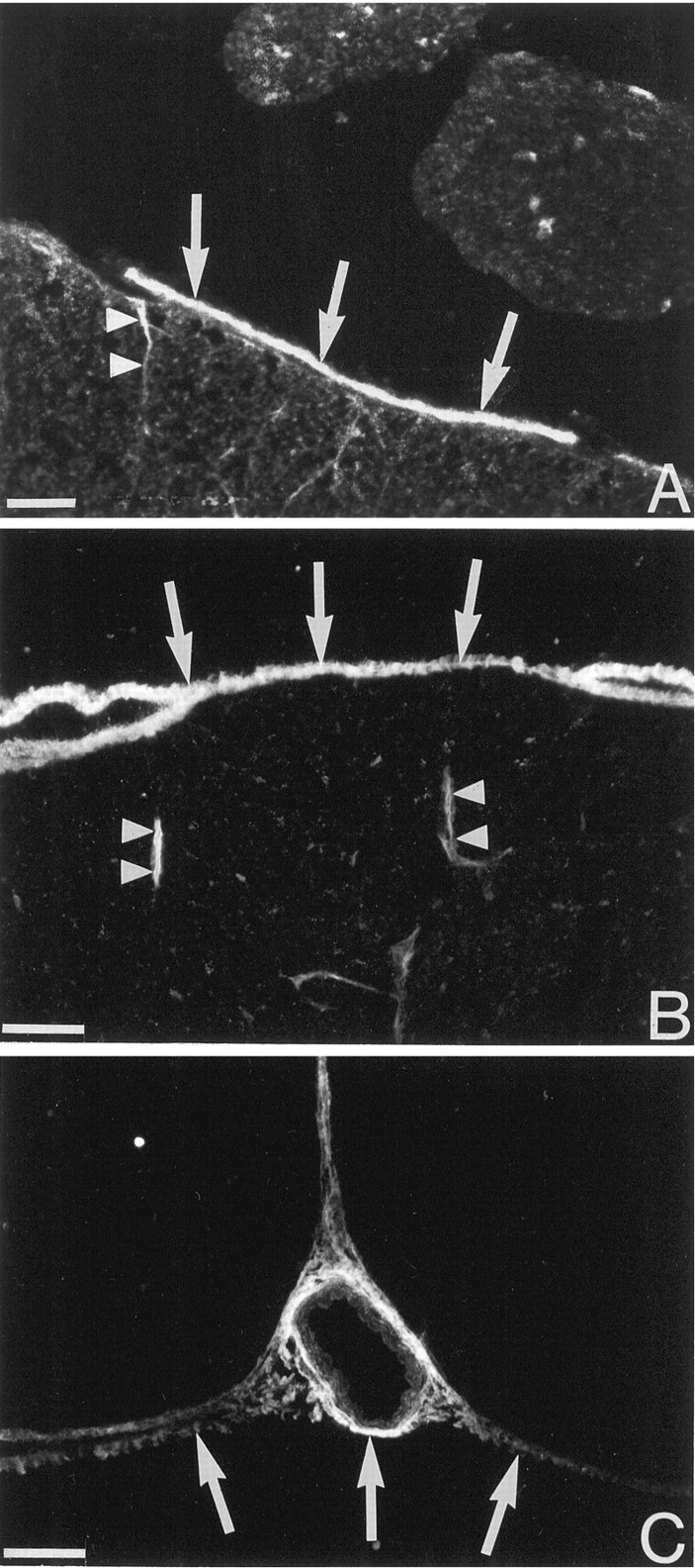
trkA-like immunoreactivity in the pia mater. The micrographs show immunohistochemical detection of trkA at the surface of the uninjured adult cat (A) and rat (B, C) lumbar spinal cord. The arrows indicate labeling in the pia mater. Thin cell process associated with blood vessels extend into the spinal cord (arrowheads). InC, trkA-LI is seen in the pia mater in the ventral fissure and in the pia mater surrounding a blood vessel. Scale bars, 50 μm.