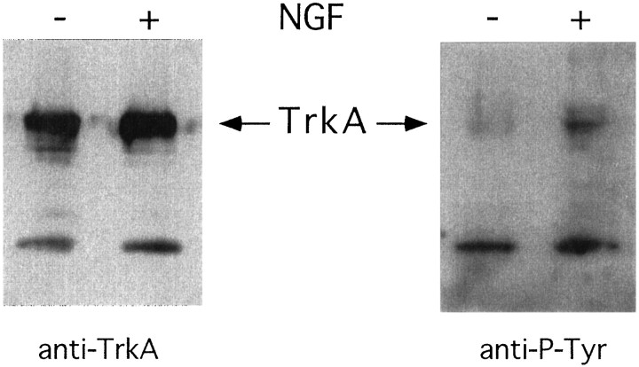
Fig. 4.
Activation of trkA by NGF in leptomeningeal cells. The cells were lysed, immunoprecipitated with trk-antiserum, separated on SDS-PAGE, transferred to a membrane, and incubated with trkA (left panel) or phosphotyrosine (right panel) antibodies. Cells were grown in the absence of NGF (−) or in the presence of NGF for 5 min before lysis (+).

