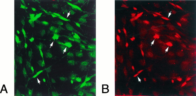
Fig. 6.
Colocalization of trkA and p75 immunoreactivity in leptomeningeal cells. Meningeal cells kept in culture for 5 d were fixed and incubated with antibodies against p75 and trkA. Fluorescent secondary antibodies were used to visualize p75 (A, FITC, green) or trkA (B, Cy3,red). There is a colocalization of p75 and trkA in most of the immunoreactive cells (arrows).

