
Fig. 3.
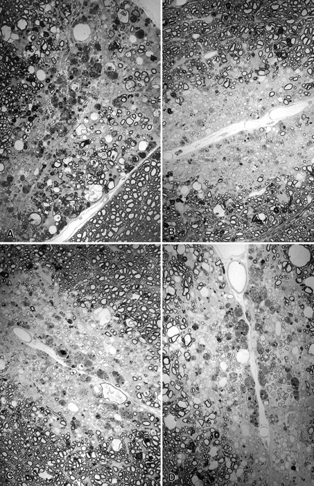
Spinal cord sections from SJL/J mice injected with lysolecithin and treated with A, PBS control;B, methylprednisolone; C, mAb 94.03; orD, polyclonal Ig directed against spinal cord homogenate (SCH/Ig). Treatment was begun on day 0 (methylprednisolone) or day 7 (PBS control, mAb 94.03 or SCH/Ig) and continued until day 21 at time of kill. Note focal area of demyelination with dense macrophage infiltration with minimal remyelination in the mouse treated with PBS control (A). In contrast, there is oligodendrocyte remyelination, characterized by abnormally thin myelin sheath relative to axon diameter, and Schwann cell remyelination in mice treated with methylprednisolone (B), mAb SCH94.03 (C) or SCH/Ig (D). The number of remyelinated axons from sections such as these are shown in Tables 1 and 2 (× 750).