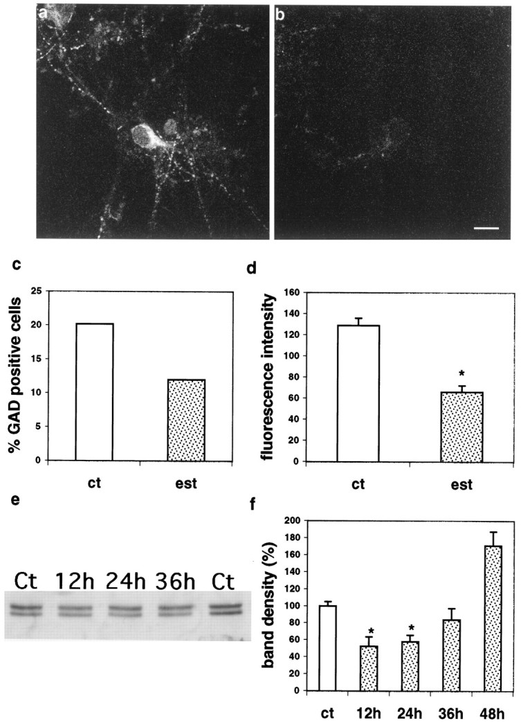
Fig. 2.

Estradiol suppresses GAD expression in cultured hippocampal neurons. A, Control. B, Twenty-four-hour exposure to estradiol. Scale bar, 20 μm.C, Percentage of GAD-positive neurons of the total cells in all of the randomly sampled fields from control (ct) and estradiol (est)-treated cultures; estradiol cultures are different from controls using χ2 analysis;p < 0.01. D, Estradiol reduces the fluorescence intensity of GAD staining (scale in arbitrary fluorescence units); mean ± SEM. E, Western blotting of GAD-stained gels, detecting both GAD65 (bottom band) and GAD67 (top band). F, Summary of densitometry analysis of effects of estradiol on total GAD immunoreactivity shown in E. Scale is percentage of matched untreated control lanes. In D andF, asterisks denote difference from controls using t tests; p < 0.01.
