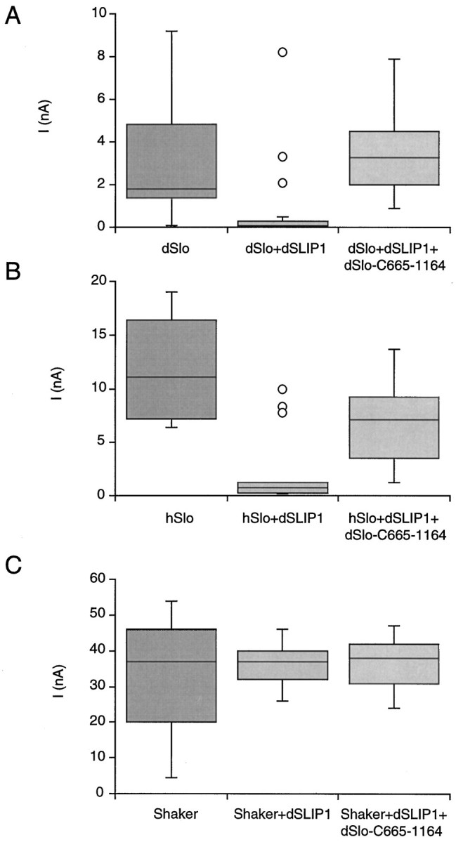
Fig. 4.

Coexpression with dSLIP1 reduces BK current amplitudes. Current amplitudes at 100 mV were determined in inside-out patches from oocytes excised into 10 μmCa2+ (A, B) or by using the two-electrode voltage clamp (C). Oocytes expressed dSlo (A), hSlo (B), or Shaker (C) channels either alone (left columns) or in combination with dSLIP1 (middle columns) or with dSLIP1 and dSlo-C665–1164 (right columns). Patch currents are shown as box plots in which the median is represented by a line separating the upper and lower quartiles (UQ, LQ). The box (interquartile distance, IQD) contains ± 25% of the data points; the error bars mark the minimum and maximum values that fall within UQ + 1.5 × IQD and LQ + 1.5 × IQD. The outliers in A and B were recorded from a single oocyte in each case. The numbers of patches in each plot were 21 for dSlo and dSlo + dSLIP1, 18 for dSlo + dSLIP1 + dSlo-C665–1164, and 15 for hSlo, hSlo + dSLIP1 and for hSlo + dSLIP1 + dSlo-C665–1164. Also examined were 11 oocytes expressing Shaker or Shaker + dSLIP1 and 6 oocytes expressing Shaker + dSLIP1 + dSlo-C665–1164.
