Fig. 2.

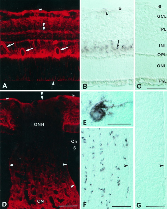
Distribution of AQP4 immunoreactivity (A, D) and AQP4 mRNA (B, E, F) in the retina and optic nerve. A, Immunofluorescence of AQP4 in the central retina. Immunolabeling extends from the inner to the outer limiting membrane (asterisk andarrowhead, respectively) and is concentrated along vessels (arrows) and the vitreal surface and in the outer plexiform layer. Note laminar labeling (double arrowhead) in the inner plexiform layer. B, Section corresponding to that in A, incubated with a digoxigenin-labeled probe to AQP4 mRNA. Note strong staining in the inner nuclear layer (arrow) and scattered and weaker staining in the nerve fiber layer (arrowhead). Interference optics. Asterisk, Inner limiting membrane.C, Sense control. GCL, Ganglion cell layer; IPL, inner plexiform layer; INL, inner nuclear layer; OPL, outer plexiform layer;ONL, outer nuclear layer; PhL, photoreceptor layer. D, The optic nerve head (ONH; longitudinal section) shows weak labeling compared with the retina and the optic nerve (ON). The choroid (Ch) and sclera (S) are immunonegative. Asterisks and double arrowhead, Vitreal surface of retina and optic nerve head, respectively; arrowheads, pial surface of optic nerve.E, F, High-magnification (E) and low-magnification (F) micrograph of AQP4 mRNA containing cells in the optic nerve. Longitudinal section, interference optics. Arrowhead, Pial surface of nerve.G, Sense control. Scale bars: A–C, 50 μm; D, F, G, 100 μm; E, 25 μm.
