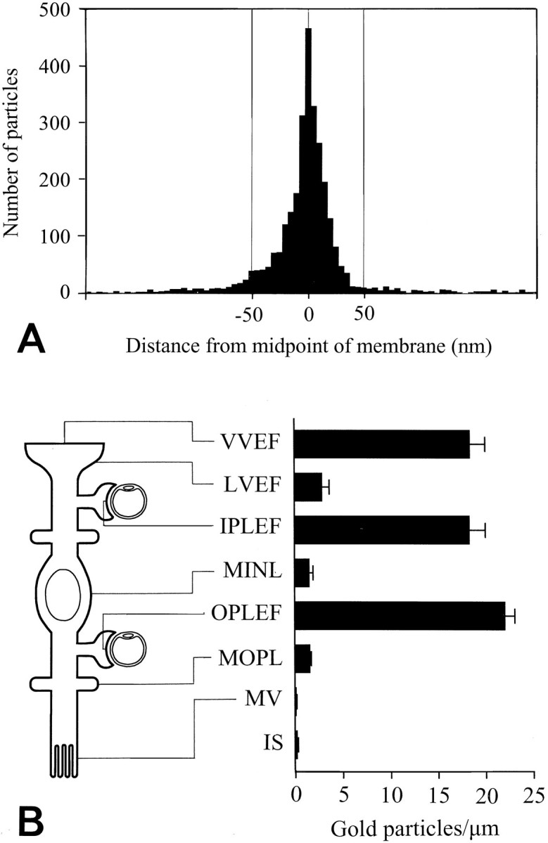
Fig. 7.

Quantitative analysis of AQP4 immunogold labeling in Müller cells. A, Distribution of gold particles along an axis perpendicular to the Müller cell plasma membrane. The ordinate indicates number of gold particles per bin (bin width, 5 nm). The data were pooled from all Müller cell membrane domains represented in B (each membrane fragment was 0.4–7 μm; the total number of gold particles was >3000). The peak coincided with the plasma membrane (0corresponds to midpoint of membrane) and the particle density approached background level at ∼50 nm from the membrane (inside negative). B, Diagram showing gold particle densities in different Müller cell membrane domains. Particles were included if they were situated within 50 nm of the membrane (cf.A). The location of each domain is indicated at theleft. VVEF, Vitreal membranes of vitreal end feet (number of observations, n = 26);LVEF, lateral membranes of vitreal end feet (n = 27); IPLEF, perivascular end feet in the inner plexiform layer (n = 17);MINL, Müller cell membranes in the inner nuclear layer (n = 14); OPLEF, perivascular end feet in the outer plexiform layer (n = 55);MOPL, Müller cell processes in the outer plexiform layer (n = 93); MV, Müller cell microvilli (n = 43). The photoreceptor inner segments (IS) are included for comparison (n = 42). Values are mean number of gold particles per micrometer ± SEM. The values for the end feet membranes (VVEF, IPLEF, and OPLEF) are significantly different from all other values (p < 0.05, Student–Newman–Keuls test). The values for microvilli and photoreceptor inner segment membranes were significantly different from LVEF, MINL, andMOPL (p < 0.05, Student–Newman–Keuls test; for statistical comparison the data from the latter three membrane domains were pooled. Membranes were concatenated to form fragments with a minimum length of 10 μm). Vitreal end feet of astrocytes also displayed a polarized expression of AQP4 (data not shown). Mean numbers of gold particles per micrometer ± SEM (n) in the vitreal and lateral membrane domains of astrocytic end feet were 12.8 ± 1.9 (16) and 0.6 ± 0.2 (12), respectively.
