
Fig. 1.
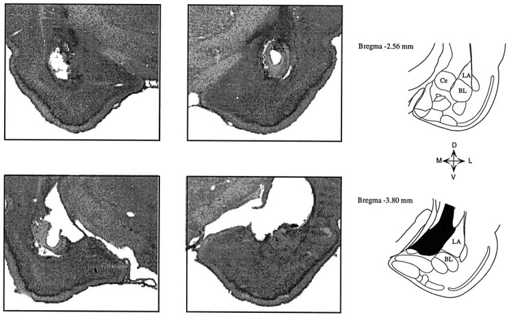
Photomicrographs of coronal sections showing a typical bilateral amygdala lesion. Lesions were centered at the lateral and basolateral nuclei, and in some cases damage extended into the central nucleus. Damage to the caudate nucleus above the amygdala was variable from case to case. Ce, Central nucleus;LA, lateral nucleus; BL, basolateral nucleus.