
Fig. 1.
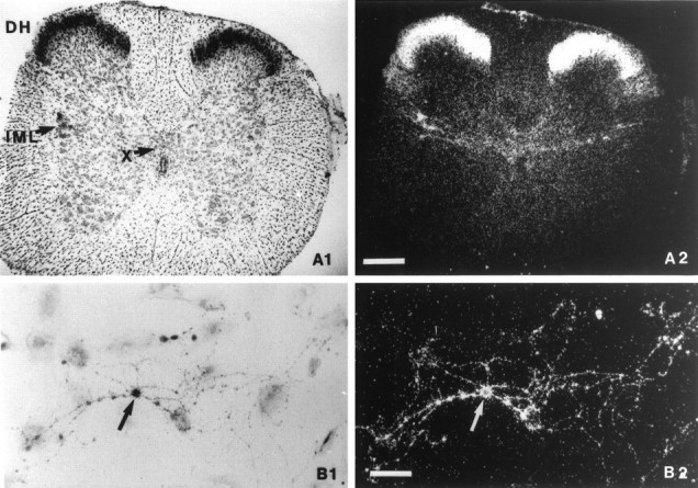
Historadioautographic detection of OT binding sites. A1, B1, Bright-field photomicrographs; A2, B2, dark-field photomicrographs. A, Transverse spinal cord section of a 15-d-old rat at the upper lumbar level showing a high density of OT binding sites in the DH, the intermediolateral cell column (IML), and lamina X (X) dorsally to the central canal. Scale bar, 250 μm. B, Culture of dissociated DH neurons after 12 d in vitro. OT binding sites were present on a cell body (arrow) and its related neurites. Scale bar, 25 μm.