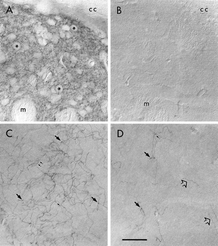
Fig. 3.

Light micrographs illustrating peroxidase immunoreactivity for DAT in the rat forebrain. A, In the dorsolateral striatum, dense peroxidase product for DAT is localized to the neuropil immediately beneath the corpus callosum (cc). Perikarya (asterisks) and bundles of myelinated axons (m) are unlabeled.B, No DAT immunoreactivity is detected in the same striatal region of sections incubated in primary antibody preadsorbed with the DAT antigen. C, In the rostral portion of the anterior cingulate cortex, a dense cluster of DAT-immunoreactive fibers is visualized in layer III. These presumed axons exhibit the branching (small arrows) and beading (large arrows) that are characteristic of terminal fibers. D, In layer VI of the prelimbic cortex from the same section as that shown inC, sparse fibers immunoreactive for DAT are observed. Although some are beaded or branched, others appear to be fibers of passage (open arrows) exiting the white matter. InA–D, up is dorsal andleft is medial. Scale bar, 150 μm.
