
Fig. 5.
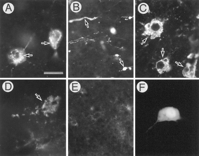
Degeneration of APP-accumulating hippocampal neurons. AxCAYAP was injected into the dorsal hippocampus. Tissues were stained with antibody AC-1 (A–E) and anti-β-gal antibody (F) on day 4 (A–D, F) and day 15 (E). The regions examined are A, C, D, F, the hilus of the dentate gyrus; B, the stratum lucidum; E, the granule cell layer of the dentate gyrus. Highly APP-immunoreactive materials are detected in the perikarya (arrows in A), dilated neurites (arrows in B), deformed intracellular membranes (arrows in C), and amorphous extracellular depositions (arrow inD). Very weak APP-immunoreactive materials are detected in the dentate gyrus on day 15 (E). Most of the β-gal-immunopositive neurons show no apparent degeneration (F). Scale bar (shown inA): A, C, D, F, 20 μm; B, E, 50 μm.