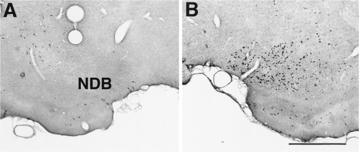
Fig. 1.
Choline acetyltransferase (ChAT) staining in the nucleus of the diagonal band (NDB). A, ChAT staining in NDB 10 d after microinjections of192-IgG-saporin unilaterally in NDB. Note the lack of ChAT-positive cells in NDB ipsilateral to the saporin lesion.B, ChAT staining in the contralateral control NDB. Scale bar, 700 μm.

