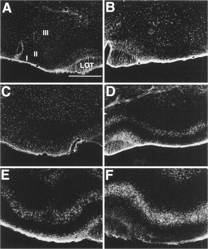
Fig. 6.

Glial fibrillary acidic acid (GFAP) staining in the piriform cortex. Sections were stained for GFAP histochemistry 10 d after unilateral lesions with 192-IgG-saporinipsilaterally in the NDB. Low levels of GFAP staining in control (no soman) animals are present in the PC ipsilateral (A) and contralateral (B) to the lesion site. By 45 min after an intramuscular injection of soman, GFAP staining is indistinguishable from controls in the PC ipsilateral to the lesion injection site (C). However, discrete layer-specific GFAP staining is present in layer II of the contralateral PC (D). By 90 min after soman, increased layer-specific increases in GFAP staining are observed in the PC ipsilateral to the lesioned NDB (E). Further increases in GFAP staining in layer II are observed in the contralateral PC (F). Scale bar, 750 μm. I, II, III, Layers of PC;LOT, lateral olfactory tract.
