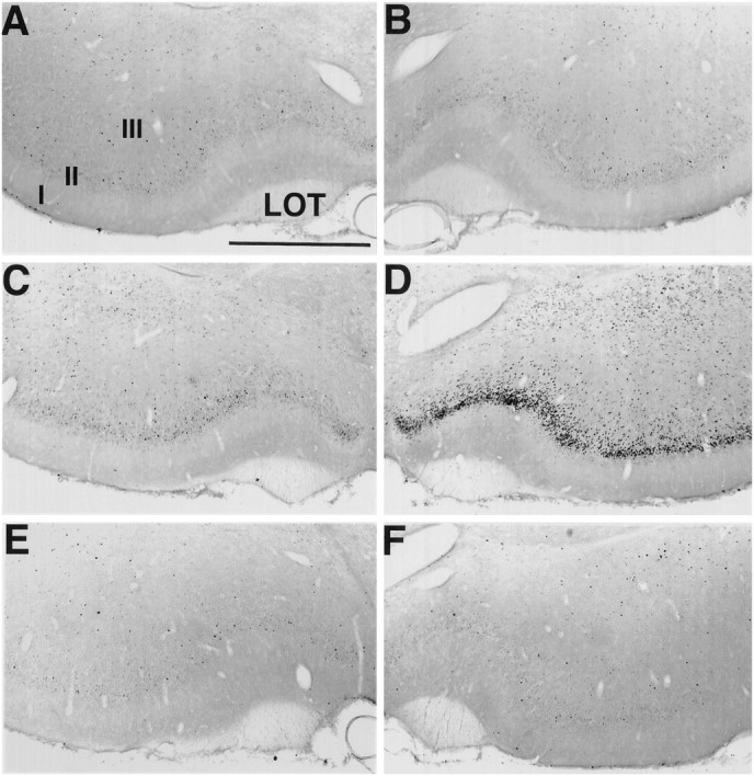
Fig. 8.

Fos staining in the piriform cortex. Sections were stained for Fos immunohistochemistry after focal ipsilateral stimulation of the NDB. Fos staining in control (no stimulation) animals is absent in the PC contralateral (A) and ipsilateral (B) to the stimulation electrode. By 45 min after focal stimulation of the NDB, Fos staining is similar to that in controls in the PC contralateral to the stimulation site (C). However, robust Fos staining is present in the PC ipsilateral to the NDB stimulation site (D). Pretreatment with the selective muscarinic receptor antagonist scopolamine inhibits increased Fos staining in the PC after 45 min of NDB stimulation (ipsilateral, in E; contralateral, in F). Scale bar, 750 μm.I, II, III, Layers of PC; LOT, lateral olfactory tract.
