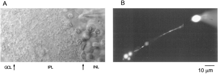
Fig. 1.
Identification of RBCs in rat retinal slices.A, Transmitted light image of a retinal slice preparation with the recording pipette placed on a cell body located in the inner nuclear layer. Arrows indicate the approximate boundaries of the different layers observed. INL, Inner nuclear layer; IPL, inner plexiform layer;GCL, ganglion cell layer. B, The same cell with its axon descending straight along the IPL and branching into three small processes in the outer part of the IPL (close to the ganglion cell layer) can be observed 10 min after breaking into the cell. A photomontage of images obtained at three different focal planes is shown. The cell was filled with 200 μm OG5. Scale bar applies to A and B.

