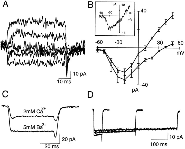
Fig. 2.
ICa in rod bipolar cells. A, Currents recorded under somatic whole-cell voltage clamp in a RBC dialyzed with CsGlu. Depolarizing pulses were applied from a holding potential of −70 mV in 20 mV increments.B, Pooled data on the current–voltage relationship obtained in RBCs dialyzed with CsGlu in 2 mm external Ca2+ (filled circles;n = 8) and in 5 mm external Ba2+ (open triangles;n = 5). Error bars indicate SEM. In both groups the activation threshold for the inward current was at −40 mV, and peak amplitudes were reached at −20 mV. Note the decrease in outward current when cells were bathed in external Ba2+.C, Comparison of the inward currents elicited by pulses to −30 mV (Vh, −70 mV) in an RBC when the slice is perfused with 2 mmCa2+ or 5 mm Ba2+. Each trace is the average of 24 individual records.D, Negligible inactivation ofICa in RBCs. Currents were elicited by depolarizing pulses to −20 mV (Vh, −70 mV) of 50, 250, and 500 msec duration. Each traceis the average of 24 records.

